Echocardiography at Wellington Hospital began in the mid 1970s with the availability of M-mode echo.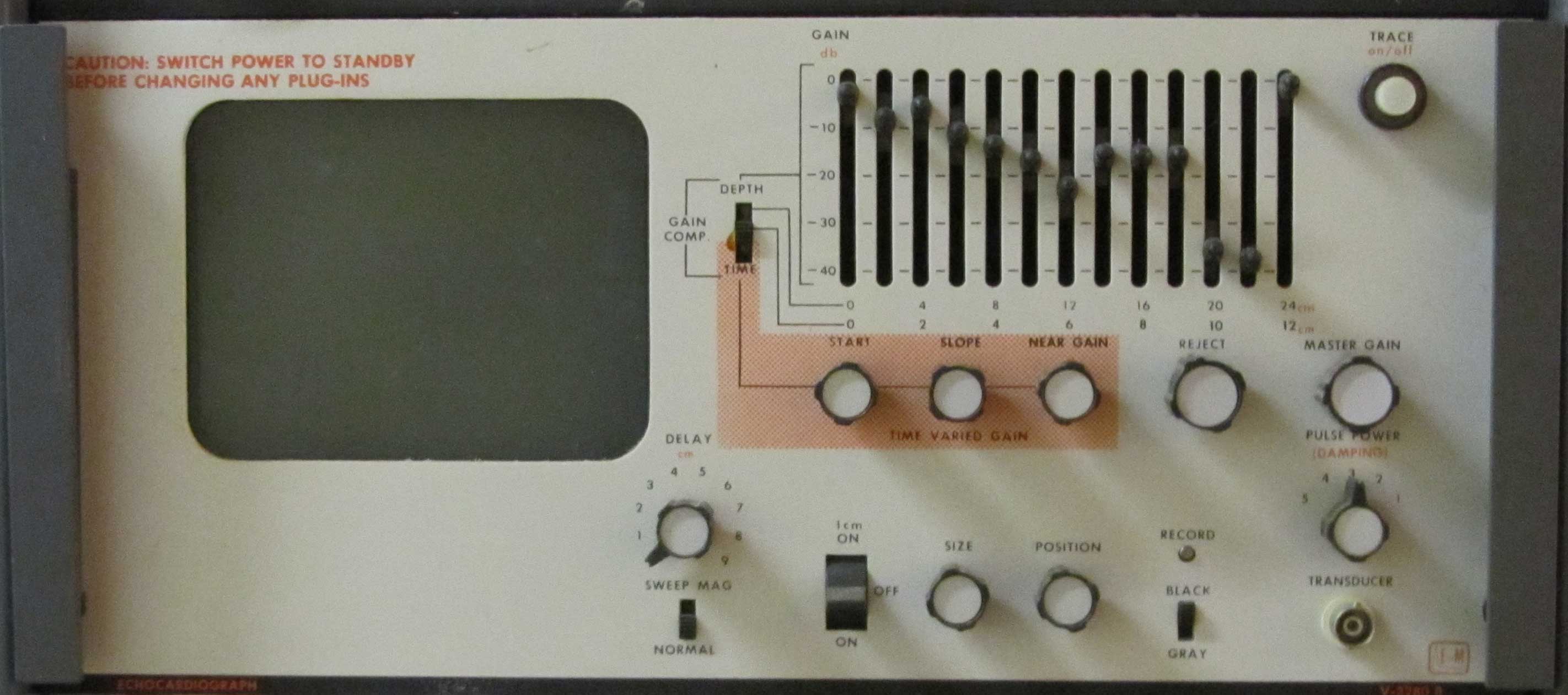
The original equipment was a module contained in a Honeywell multichannel recorder. It had a tiny screen on which to view the M-mode tracings.
A little later we acquired a Picker Echoview M-mode echocardiograph. This was a stand-alone piece of equipment and in time became useful in the assessment of mitral stenosis, LV function and pericardial effusion.
Although there was some value in having this 'primitive' technology, it wasn't until 1983 that the Department acquired its first 2D echo machine, and now there was something to get excited about. The bulk of the studies in the early years were done by David McHaffie, Ron Easthope and Vikki D'Ath who all learned on the job. Sue Pankhurst, with some echo experience gained in Australia, joined the echo team when she returned to Wellington in 1986.
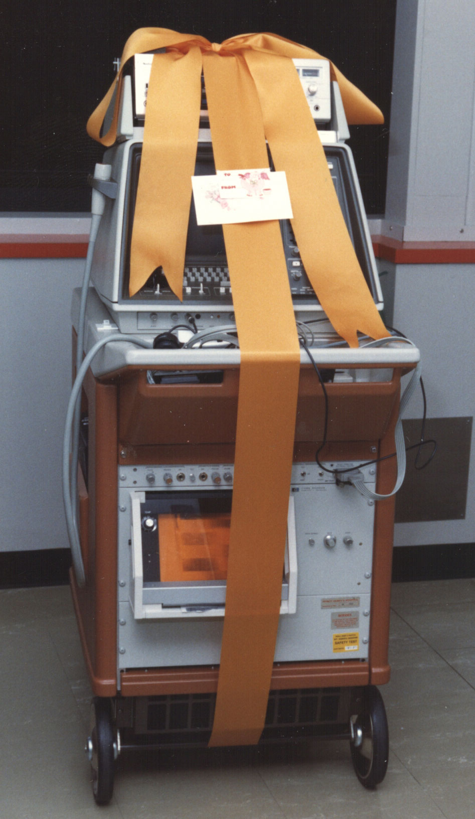
The initial equipment did not come with colour doppler. That arrived with the purchase of a HP Sonos 1000 machine in 1987, and this came gift-wrappped! This was the first colour doppler machine in the country.
From a modest total of around 1000 studies done in 1985, the numbers had trebled by 1995.
The equipment came with a paper print-out module which proved to be of little use. When we made this purchase, advice received suggested that we opt for the Betamax format tape deck, seen here mounted on the top of the machine.
It wouldn't be too many years later that this video format became obsolete.
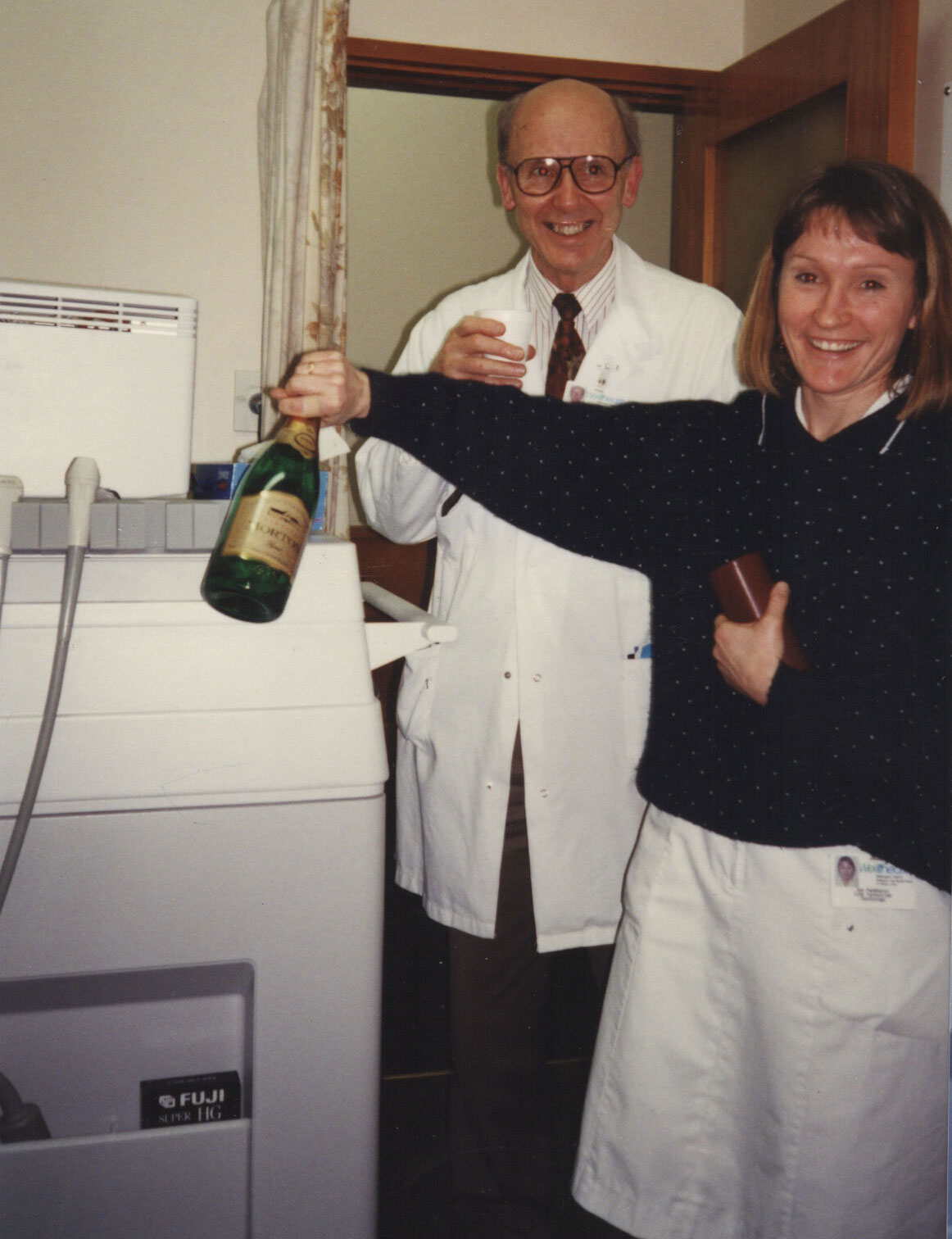
In October 1994 the department took delivery of one HP 2000 and two HP 2500 echo machines. Ron and Sue clearly thought there was much to celebrate.
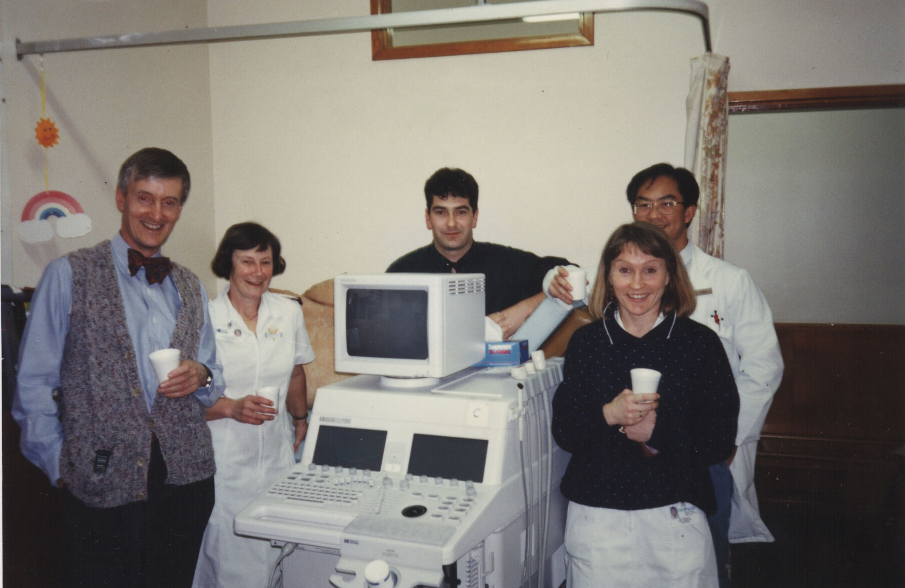
In the photograph below are, from left, David McHaffie, Hazel Milliken, Nigel Lever, Sue Pankhurst and behind Sue, Geoffrey Chia.
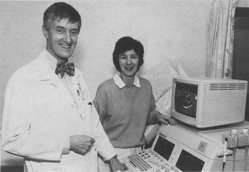
The photograph above shows David McHaffie and Carol Allan with one of the HP 2500 machines. Included in the purchase was the hospital's first multiplane transoesophageal echo probe. The cost of the total package was $700,000.
Later, having received a generous donation from a family who had a child with congenital heart disease, a special paediatric echo probe was purchased.
The HP 2000 was kept in the Cardiac Care Unit giving easy access for studies on cardiac inpatients.
The introduction of echocardiography caught the imagination of David McHaffie and Ron Easthope who adopted a hands-on approach.
Considerable experience was acquired, and credit must go to a number of technicians who quickly became skilled in the technique. Stats available to 1996 reveal:
| name | period | # studies |
|---|
| Vikki D'Ath | 1984-1990 | 3,077 |
| Sue Pankhurst | 1986-1994 | 6,651 |
| Carol Allan | 1988-1995 | 4,252 |
| Helen Templeton | 1987-1996 | 2,241 |
| Hazel Milliken | 1991-1996 | 2,958 |
Other notable contributors, for whom numbers are not available, include Sue Winter and Sally Cadacio. Total study numbers for 1985-1995 appear below:

The total number of echos done during the financial year 2003/2004 was 4,022.
Arguably, the Wellington Hospital team became the leaders in echo experience in NZ. Over the period 1988 - 1994 we hosted a number of echo symposia, attracting registrants from around the country. The largest of these meetings was held in 1994. Another major symposium was organised by Alex Sasse in 2012.
I developed an interest in foetal echocardiography. Working alongside Peter Stone and Sarath Weerasinghe (Obstetricans) and Jenny Flower (Sonographer) I gradually gained experience in recognising important foetal heart defects.
In 1990, I spent a week working with Dr Lindsey Allan at Guys Hospital in London, she being the UK's foremost expert in this field.
With the arrival of the multiplane transoesophageal probe in 1994, a new demand for echo services developed. The cardiac surgeons, notably Allan Hilless and Ashok Sharma, liked to have transoesophageal echos done intraoperatively, before and after mitral valve repair particularly. Ron and David assisted whenever they could.
Cardiologists were relieved of this 'chore' when the anaesthetists acquired their own echo machine and TOE probe and received training in the technique. The anaesthetists have now gained an edge in having the hospital's only 3-D TOE probe.
New echo equipment was purchased in 2000. Our traditional allegiance to the HP stable was broken. One Vinmed Vivid 5 and two Vivid 3 machines were installed. The following year the Vinmed digital archiving system was acquired.
No longer was there a reliance on the use of videotapes, and it was now possible to undertake remote post-processing and reporting. In addition, cardiologists seeing clinic patients could now view studies away from the echocardiography machine rather than interrupting a subsequent patient study.
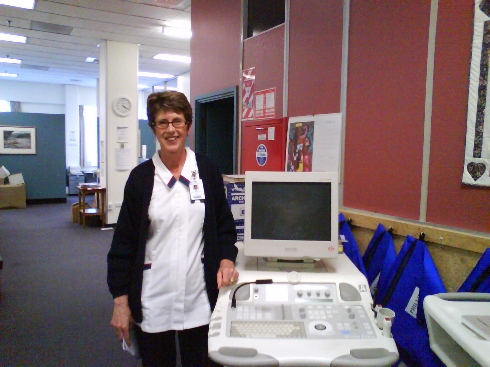 Sue Winter with the Vivid 5 machine in the temporary cardiology location in the CSB.
In 2007, Alexander Sasse was appointed to the staff and brought with him significant imaging experience.
The following year four new echo machines were purchased. Two Vivid 7, one S6, and one portable Vivid I 'laptop' machine made up the new suite from GE. The two Vivid 3 machines were retained but seldom used as they required scans to be loaded on to disc for transfer.
In 2010, a workstation was installed at Kenepuru Hospital where studies were done weekly on both clinic and inpatients. Scans were able to be transferred directly to the Wellington Hospital archive and viewing room.
The acquisition by Wellington of the Vinmed machines in 2000 was matched by the purchase of similar equipment at Hutt Hospital, and in time, it became possible to view Hutt studies directly in Wellington.

 Healthline
 GP
GP Afterhours
 Hospital ED